Personalized Medical Care & Dedicated International Patient Concierge


Pregnancy Care · Needs Clinic Confirmation
Fetal Ultrasound & Anomaly Screening
Fetal ultrasound is a fundamental and indispensable component of contemporary prenatal care, enabling real-time visualization of fetal development through high-frequency sound wave imaging technology.
Schedule a ConsultationComprehensive Understanding of Fetal Ultrasound in Modern Prenatal Medicine

Fetal ultrasound is a fundamental and indispensable component of contemporary prenatal care, enabling real-time visualization of fetal development through high-frequency sound wave imaging technology. This non-invasive diagnostic tool allows clinicians to assess fetal growth, structural development, organ formation, placental function, and amniotic environment without exposing the mother or fetus to radiation or invasive procedures.
The primary goal of fetal ultrasound is to ensure the healthy progression of pregnancy while identifying potential abnormalities at the earliest possible stage. Early detection is essential because many congenital conditions or developmental variations are most effectively managed when identified before birth.
At THE YONSEI MIZ WOMEN'S CLINIC, fetal ultrasound is not performed as an isolated procedure but as part of an integrated prenatal care system. Each examination is interpreted by experienced obstetric specialists who analyze findings in relation to maternal history, gestational age, genetic risk factors, and overall pregnancy progression.
This comprehensive approach ensures not only diagnostic accuracy but also reassurance and clarity for expecting parents throughout the pregnancy journey.
Stages and Types of Fetal Ultrasound Evaluation
Fetal ultrasound is performed at multiple standardized stages of pregnancy, each designed to evaluate specific aspects of fetal development and maternal-fetal health.
First-Trimester Comprehensive Assessment
The early pregnancy scan confirms intrauterine pregnancy, fetal heartbeat, gestational sac location, and accurate gestational dating. It is essential for establishing baseline pregnancy viability and identifying early complications such as ectopic pregnancy or early developmental concerns.
This stage also provides the foundation for subsequent prenatal monitoring and risk assessment.
Nuchal Translucency (NT) and Early Risk Screening
Conducted typically between 11–14 weeks of gestation, NT screening measures the fluid accumulation at the posterior aspect of the fetal neck. This measurement is an important marker used in early assessment of chromosomal abnormality risk.
When combined with maternal serum screening tests, it contributes to a more comprehensive evaluation of early fetal development and genetic risk stratification.
Second-Trimester Detailed Anomaly Scan
The mid-pregnancy anomaly scan, usually performed between 18–24 weeks, is one of the most critical stages of fetal evaluation. It involves a systematic and detailed examination of fetal anatomy.
Structures evaluated include the brain, face, spine, heart, lungs, diaphragm, abdominal organs, kidneys, bladder, extremities, and skeletal development. Placental position, umbilical cord structure, and amniotic fluid volume are also assessed in detail.
This scan plays a key role in identifying congenital structural abnormalities and guiding further prenatal management if required.
Fetal Growth and Biophysical Profile Assessment
Serial growth scans monitor fetal size, estimated weight, and proportional development over time. These measurements are compared against standardized growth curves to ensure appropriate fetal development.
The biophysical profile may also include assessment of fetal movements, tone, breathing patterns, and amniotic fluid levels, providing a broader evaluation of fetal well-being.
Doppler Ultrasound and Placental Circulation Analysis
Doppler studies evaluate blood flow within the uterine arteries, umbilical artery, and fetal circulation. This allows assessment of oxygen and nutrient delivery between mother and fetus.
Abnormal flow patterns may indicate placental insufficiency or fetal growth restriction and require closer monitoring or intervention.
Meet the Pregnancy Care Specialists
 |
 |
|---|---|
| **Dr. Jong-sik Jeon, MD | |
| Representative Director** | |
| Specialist in Obstetrics & Gynecology | **Dr. Ye-ri Lee, MD |
| Vice Director** | |
| Specialist in Obstetrics & Gynecology |
THE YONSEI MIZ WOMEN'S CLINIC is led by highly experienced board-certified obstetrician-gynecologists specializing in fetal imaging, prenatal diagnosis, and maternal-fetal medicine.
Dr. Jong-sik Jeon
Dr. Jong-sik Jeon is the Chief Director of THE YONSEI MIZ WOMEN'S CLINIC and a board-certified obstetrician-gynecologist. He graduated from Yonsei University College of Medicine and has accumulated over 30 years of clinical experience in obstetrics and gynecology.
He has performed more than 30,000 gynecologic surgeries and procedures. His past leadership roles include Director of Gangnam MizMedi Hospital, Medical Director of Gangnam Major Hospital, Clinical Professor at Yonsei University College of Medicine, and Clinical Professor at Yonsei University Gangnam Severance Hospital.
His expertise includes high-risk pregnancy management, fetal ultrasound interpretation, prenatal diagnosis, and comprehensive obstetric care.
Dr. Ye-ri Lee
Dr. Ye-ri Lee serves as Vice Director of THE YONSEI MIZ WOMEN'S CLINIC. She graduated from Kyung Hee University College of Medicine and completed obstetrics and gynecology residency training at Samsung Seoul Hospital.
She previously served as Director of the Department of Obstetrics and Gynecology at Kwak Women’s Hospital and Adjunct Professor at Samsung Seoul Hospital. Her clinical expertise includes prenatal care, fetal development monitoring, and maternal-fetal health management.
Together, the specialists provide highly precise, evidence-based fetal ultrasound and anomaly screening with a strong emphasis on diagnostic accuracy, safety, and maternal reassurance.
What Fetal Ultrasound Can Detect and Evaluate
Detailed Fetal Organ Development
Ultrasound enables visualization of fetal brain development, cardiac structure, pulmonary formation, renal development, and gastrointestinal anatomy.
Structural Congenital Abnormalities
Early identification of structural anomalies allows timely counseling, further diagnostic evaluation, and appropriate pregnancy management planning.
Growth Restriction or Excessive Growth Patterns
Serial measurements help identify fetal growth restriction or macrosomia, allowing adjustments in prenatal care strategy.
Placental Health and Function
Placental position, morphology, and function are assessed to ensure adequate support for fetal development.
Amniotic Fluid Balance
Amniotic fluid levels are monitored as an important indicator of fetal well-being and uterine environment stability.
Step-by-Step Fetal Ultrasound Process

1. Pre-Examination Clinical Review
Maternal history, gestational age, and prior ultrasound results are reviewed before imaging begins.
2. High-Resolution Imaging Acquisition
Advanced ultrasound equipment is used to obtain detailed fetal and uterine images.
3. Systematic Anatomical Survey
Each fetal organ system is evaluated in a structured and standardized manner.
4. Doppler and Growth Analysis
Blood flow patterns and fetal growth parameters are carefully measured and interpreted.
5. Specialist Interpretation and Reporting
Findings are analyzed and explained clearly, with emphasis on clinical significance.
6. Personalized Follow-Up Planning
A structured prenatal monitoring plan is developed based on findings and pregnancy stage.
Safety, Accuracy, and Clinical Importance
Fetal ultrasound is a completely safe, non-invasive imaging method that uses sound waves and does not involve radiation exposure. It is widely recognized as a standard and essential component of prenatal care.
Most fetal ultrasound examinations confirm normal development and provide reassurance to expectant parents. When abnormalities are detected, early identification enables timely intervention, counseling, and management planning.
Emotional Reassurance and Maternal Experience
Beyond its diagnostic value, fetal ultrasound provides significant emotional reassurance during pregnancy. Visualizing fetal development and receiving expert interpretation helps strengthen maternal bonding and reduce anxiety.
At THE YONSEI MIZ WOMEN'S CLINIC, communication is delivered in a clear, compassionate, and supportive manner to ensure that patients fully understand their pregnancy progress.
Why Choose THE YONSEI MIZ WOMEN'S CLINIC?
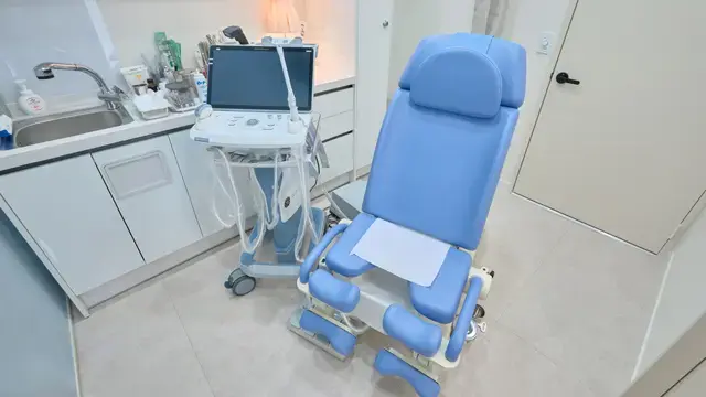
THE YONSEI MIZ WOMEN'S CLINIC is a dedicated women’s healthcare center specializing exclusively in obstetrics and gynecology, with advanced expertise in pregnancy care and prenatal diagnostics.
Patients choose the clinic for:
- Advanced fetal ultrasound imaging systems
- Highly experienced OB-GYN specialists in prenatal diagnosis
- Comprehensive multi-stage anomaly screening programs
- Structured fetal growth and development monitoring
- Early detection of congenital abnormalities
- Safe, non-invasive pregnancy evaluation methods
- Clear and detailed clinical interpretation
- Compassionate, patient-centered obstetric care
Each examination is performed with clinical precision, diagnostic excellence, and a strong focus on maternal and fetal well-being.
Schedule Your Fetal Ultrasound & Anomaly Screening
If you are pregnant and require routine fetal monitoring, anomaly screening, or detailed prenatal evaluation, THE YONSEI MIZ WOMEN'S CLINIC provides advanced fetal ultrasound services and comprehensive obstetric care. Our experienced specialists ensure accurate diagnosis, clear communication, and continuous pregnancy support in a safe and reassuring clinical environment.
Book Your Fetal Ultrasound Appointment Today
Contact THE YONSEI MIZ WOMEN'S CLINIC to schedule expert prenatal imaging and comprehensive pregnancy monitoring care.
Other Services
Explore additional treatments offered by our specialists.

Colposcopy
Colposcopy is an advanced diagnostic gynecologic procedure that allows detailed examination of the cervix, vagina, and vulvar tissues using a specialized magnifying instrument known as a colposcope.
Learn More

PCOS — Adolescent Care
Polycystic Ovary Syndrome (PCOS) is one of the most common endocrine disorders affecting adolescent and young women worldwide.
Learn More

Vaginal Dryness & Relaxation Treatment
Vaginal dryness and relaxation concerns are commonly associated with hormonal changes during perimenopause and menopause.
Learn More